Confocal Imaging
Confocal Fluorescence Imaging
Image Analysis
Training
Experimental Design
The confocal digital imaging unit is located in Room H1710 in the Health Sciences Centre.
The unit operates on a fee-for-service basis, and is available to both internal and external users.
Hours of Operation
Monday through Friday
9:00am-5:00pm. (Fall & Winter)
9:00am-4:30pm (Summer)
Equipment and Service
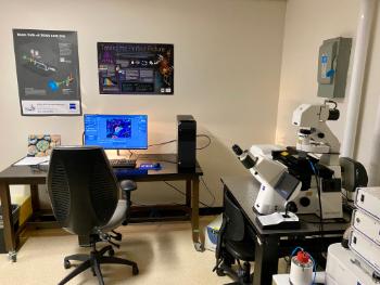
Zeiss LSM 900 with Airyscan 2
-488 nm -561 nm -640 nm
|
 |
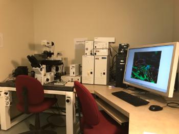
Olympus Fluoview FV1000 laser scanning microscope
- 488 nm Argon - 515 nm Multi Argon - 543 nm Green HeNe - 568 nm Krypton/Argon - 633 nm Red HeNe
|
 |
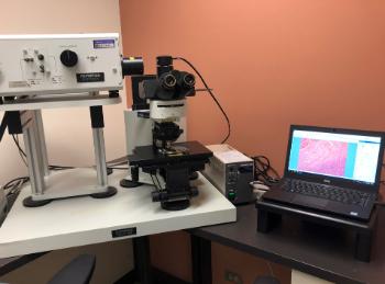
Olympus Fluoview FV300 laser scanning microscope
|
 |
Scheduling and Data Policies
- Booking time is through the scheduler located in Room H1710, or via email (see below).
- A requisition is required at time of use.
- Users are responsible for removing data files at time of acquisition
Biosafety Guidelines
For all biosafety guidelines refer to Memorial University Biosafety Policies and Procedures Manual.
All samples transported to the laboratory must be in a sealed container according to biosafety
guidelines. Laboratory coats and PPE must be worn at all times.
Please Note: Users must add room H1710 to their current Biosafety certificate to use the facility.
Acknowledgements
Please remember to acknowledge any contributions of the EFCF in your publications, presentations, posters, and grant applications.
If the staff have provided technical and scientific advice beyond the routine, please consider them for authorship in your publications if appropriate. Otherwise, a simple recognition in the acknowledgments sections such as the following is appreciated: “We thank Memorial University’s Medical Laboratories-Electron Microscopy/Flow Cytometry/ Confocal Microscopy for their support with this work.”
Educational Resources
Zeiss
Olympus
Leica
Nikon
Ibidi
Mattek
Spectra of Fluorochromes
FluoroFinder
BioLegend
ThermoFisher
Mailing Address:
Confocal Imaging Unit
Room H1710
Medical Laboratories
Faculty of Medicine
300 Prince Philip Drive
St. John’s, NL, A1B 3V6
Canada
Contact Persons:
| To book instrument time or to discuss technical questions email: |
Electron Microscopy/ Flow Cytometry/Confocal Microscopy Supervisor | Medical Laboratories

Medical Technologist | Medical Laboratories
Medical Technologist | Medical Laboratories