

All text material ©2005 by Steven M. Carr
Most textbook illustrations of the Meselson - Stahl experiment show it being conducted in a conventional swinging-bucket type centrifuge rotor. Such rotors are described as preparative, because the ordinary purpose is to prepare cell fractions based on differences in density, typically by sedimenting out a heavier fraction from a lighter supernatant fraction.
The Meselson - Stahl experiment actually
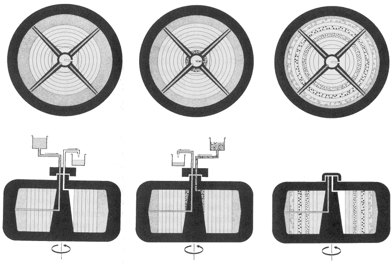
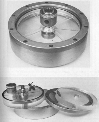
employed a less familiar type of centrifugation, which uses a zonal
analytical rotor (below, left). The circular rotor is divided into
a set of pie-shaped sectors (zones). The density-gradient medium is pumped
into the sectors while the rotor is turning at low speed. At high speed,
a gradient forms with the same density at the same radius, which thus consists
of a series of concentric rings. Once the gradient is formed, molecules
or organelles introduced into the rotor through the hub will migrate to
the radius where they have the same density as the gradient. The distribution
in the gradient can be photographed during the run through a transparent
window or aperture slot in the rotor lid, which is illuminated from below
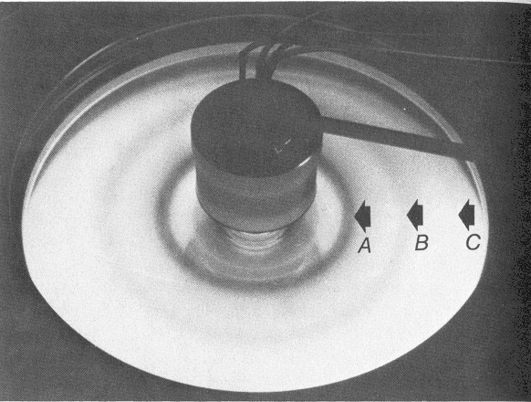
by a light source. The photograph (below, right) shows an analytical
separation of three cell components in a sucrose gradient, corresponding
to (A) microsomes, (B) mitochondria, and (C) nuclei and membranes. In a
DNA / cesium chloride gradient experiment, a uniform solution of CsCl molecules
under high centrifugal force will dissociate and the heavy Cs+
atoms will be forced towards the outside of the rotor, thus forming
a shallow density gradient. Photographs are taken with a UV light source at a fixed point on the radius:
the DNA will absorb the light, producing a dark band on a photograph, as
seen in the Meselson-Stahl results.
.


