Quatenary Structure


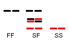
Some enzyme proteins are made up of two or more subunits that bind together to produce the functional enzyme. This quaternary structure determines the number of electrophoretic bands expected in heterozygote individuals.


Monomeric proteins – one subunit

 
Heterozygotes show two bands, one for each allozyme.
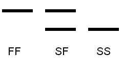


                   Electrophoresis_monomer.jpg


Dimeric proteins – two subunits


Heterozygotes shows three bands, one for each FF or SS allozyme dimer and one for the FS dimer. This heterodimer is expected to have an intermediate mobility between F and S. The dimer combinations will occur in a 1:2:1 ratio, thus the middle band should be most intense.


              Electrophoresis_dimer.jpg


Gels:


1. Monomeric enzyme protein (PGM): two heterozygotes FS and MS:


                PGM_monomer.jpg


2. Dimeric enzyme protein (PGI): SF, SS, SF
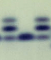
 

               PGI_dimer.jpg


3. Triploid pattern (3 allozymes: S, M, F) in a monomeric enzyme (PGM)
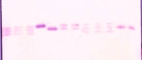
 

            PGI_triploid.jpg


Exercise:


     Use ImageJ to quantify variation in banding intensity among allozyme bands for the three gels shown above.

 
Click HERE to start the exercise.


From a lab exercise orginally prepared ©2005 by DJ Innes; text ©2008 by Steven M. Carr