Analysis of multi-locus DNA "fingerprints"
Analysis of multi-locus DNA "fingerprints"
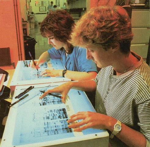
Analysis of microsatellite variation involves separation of DNA fragments by size (molecular weight) on a solid support (gel) in an electric field (electrophoresis). If the DNA fragments have been radioactively labelled (with 32P or 35S) during the amplification process, exposure of the gel to blue X-ray film produces an autoradiogram, in which each DNA fragment is seen as a black band on a blue background, as seen above. Analysis of multiple microsatellite loci on a single autoradiogram produces a complex pattern of bands (a "fingerprint") that resembles a supermarket "barcode". These geneticists are calculating sizes of fragments and comparing patterns among individuals. Modern automated DNA sequencers avoid the use of radioactive labels and can perform such calculations & comparisons automatically.